Compound Microscope Parts

A high power or compound microscope achieves higher levels of magnification than a stereo or low power microscope. It is used to view smaller specimens such as cell structures which cannot be seen at lower levels of magnification. Essentially, a compound microscope consists of structural and optical components. However, within these two basic systems, there are some essential components that every microscopist should know and understand. These key microscope parts are illustrated and explained below.
STRUCTURAL COMPONENTS
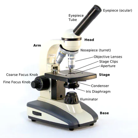
The three basic, structural components of a compound microscope are the head, base and arm.
- Head/Body houses the optical parts in the upper part of the microscope
- Base of the microscope supports the microscope and houses the illuminator
- Arm connects to the base and supports the microscope head. It is also used to carry the microscope.
When carrying a compound microscope always take care to lift it by both the arm and base, simultaneously.
OPTICAL COMPONENTS
There are two optical systems in a compound microscope: Eyepiece Lenses and Objective Lenses:
Eyepiece or Ocular is what you look through at the top of the microscope. Typically, standard eyepieces have a magnifying power of 10x. Optional eyepieces of varying powers are available, typically from 5x-30x.
Eyepiece Tube holds the eyepieces in place above the objective lens. Binocular microscope heads typically incorporate a diopter adjustment ring that allows for the possible inconsistencies of our eyesight in one or both eyes. The monocular (single eye usage) microscope does not need a diopter. Binocular microscopes also swivel (Interpupillary Adjustment) to allow for different distances between the eyes of different individuals.
Objective Lenses are the primary optical lenses on a microscope. They range from 4x-100x and typically, include, three, four or five on lens on most microscopes. Objectives can be forward or rear-facing.
Nosepiece houses the objectives. The objectives are exposed and are mounted on a rotating turret so that different objectives can be conveniently selected. Standard objectives include 4x, 10x, 40x and 100x although different power objectives are available.
Coarse and Fine Focus knobs are used to focus the microscope. Increasingly, they are coaxial knobs - that is to say they are built on the same axis with the fine focus knob on the outside. Coaxial focus knobs are more convenient since the viewer does not have to grope for a different knob.
Stage is where the specimen to be viewed is placed. A mechanical stage is used when working at higher magnifications where delicate movements of the specimen slide are required.
Stage Clips are used when there is no mechanical stage. The viewer is required to move the slide manually to view different sections of the specimen.
Aperture is the hole in the stage through which the base (transmitted) light reaches the stage.
Illuminator is the light source for a microscope, typically located in the base of the microscope. Most light microscopes use low voltage, halogen bulbs with continuous variable lighting control located within the base.
Condenser is used to collect and focus the light from the illuminator on to the specimen. It is located under the stage often in conjunction with an iris diaphragm.
Iris Diaphragm controls the amount of light reaching the specimen. It is located above the condenser and below the stage. Most high quality microscopes include an Abbe condenser with an iris diaphragm. Combined, they control both the focus and quantity of light applied to the specimen.
Condenser Focus Knob moves the condenser up or down to control the lighting focus on the specimen.