Slide Mount Instructions

Before you start building your slides, make sure you have everything you will need, including slides, cover slips, droppers or pipets and any chemicals or stains you plan to use.
You will be using two main types of slides, 1) the common flat glass slide, and 2) the depression or well slides. Well slides have a small well, or indentation, in the center to hold a drop of water or liquid substance. They are more expensive and usually used without a cover slip.
Standard slides can be either plastic or glass and are 1 x 3 inches (25 x 75 mm) in size and 1 to 1.2 mm thick.
Wet slides will use a cover slip or cover glass, a very thin square piece of glass (or plastic) that is placed over the sample drop. Without the cover in place, surface tension would cause the droplet to bunch up in a dome. The cover breaks this tension, flattening the sample and allowing very close inspection with minimal focusing. The cover also serves to protect the objective lens from interfering with the sample drop.
MOUNTS
There are four common ways to mount a microscope slide as described below:
Dry Mount
In a dry mount, the specimen is placed directly on the slide. A cover slip may be used to keep the specimen in place and to help protect the objective lens. Dry mounts are suitable for specimens such as samples of pollen, hair, feathers or plant materials.
Wet Mount
In a wet mount, a drop of water is used to suspend the specimen between the slide and cover slip. Place a sample on the slide. Using a pipette, place a drop of water on the specimen. Then place on edge of the cover slip over the sample and carefully lower the cover slip into place using a toothpick or equivalent. This method will help prevent air bubbles from being trapped under the cover slip.
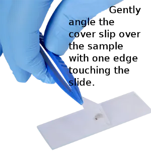
Your objective is to have sufficient water to fill the space between cover slip and slide. If there is too much water, the cover slip will slide around. Take a piece of paper towel and hold it close to one edge of the cover slip. This will draw out some water. If too dry, add a drop of water beside the cover slip. Practice this until you get used to it.

Wet mounts are suitable for studying water-bound organisms such as paramecium or bodily fluids such as saliva, blood and urine.
Section Mount
In a section mount, an extremely thin cross-section of a specimen is used. Using a microtome, cut a thin slice of your selected specimen such as an onion, and carefully set it on your slide. Then follow the instructions for a dry or wet mount. A stain can often be applied directly to the specimen before covering with a cover slip.
Section mounts are suitable for useful for a wide variety of samples such as fruit, vegetables and other solids that can be cut into small slices.
Smear
A smear is made by carefully smearing a thin layer of the specimen across a slide and then applying a cover slip. Typically, a smear should be allowed to air dry before applying a stain.
STAINS
Stains are used to help identify different types of cells using light microscopes. They give the image more contrast and allow cells to be classified according to their shape (morphology). By using a variety of different stains, you can selectively stain different areas such as a cell wall, nucleus, or the entire cell. Stains can also help differentiate between living or dead cells.
Stains tend to be grouped as neutral, acidic or basic, depending upon their chemical makeup and will attract or repel different organisms accordingly. For example, scientists and health professionals use Methylene Blue, a slightly alkaline stain, to reveal the presence of deoxyribonucleic acid, more commonly known as DNA.
Stain Types
Iodine is one of the more commonly available stains and is used to identify starch in a variety of samples. It will stain carbohydrates in plants and animal specimens brown or blue-black. Glycogen will show as red.
Methylene Blue is an alkaline stain useful in identifying acidic cell nuclei and DNA in animal, bacteria or blood samples. It’s also useful in aquariums to prevent the spread of fungal infections in fish. See more details >
Eosin Y is an acidic stain which stains pink for alkaline cells (cytoplasm, for example). It colors red for blood cells, cytoplasm and cell membranes. Eosin's most important medical uses are in blood and bone-marrow testing, including the PAP smear. See more details >
Gram's Stain is one of the most frequently used processes in identifying bacteria – used daily in hospitals. It is a primary test that quickly and cost effectively divides bacteria into one of two types: Gram positive or Gram negative. See more details >
STAINING STEPS
- Prepare a wet mount slide.
- Collect a drop of stain with an eye dropper or pipette.
- Put a drop of stain on an outer edge of your cover slide.
- Place a piece of napkin or paper towel against the opposite side of your cover slip, right up against the edge. This will help draw the stain under the cover and across the specimen.
- You may need to add another drop to ensure complete coverage.
- The slide is now ready for viewing.